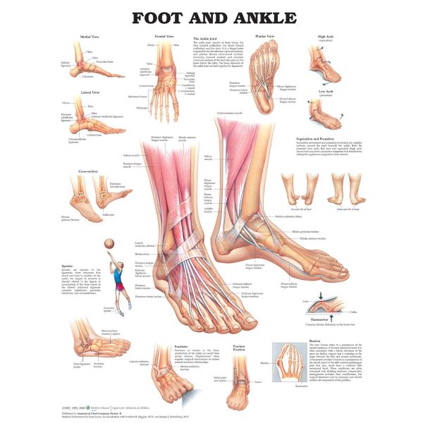
Foot & Ankle Laminated Poster
The large central figure shows normal foot and ankle anatomy including bones, muscles and tendons.
Smaller illustrations show the following details:
- medial and lateral view of the bones of the foot and ankle
- frontal view of the bones of the foot and ankle
- plantar views of the foot
- cross section of the ankle joint showing extension and flexion
Common injuries and problems are also illustrated and explained:
supination and pronation
hammertoe
bunion
sprains
fractures
fracture fixation.
Dimensions for this chart are 20″ x 26″ (51cm x 66cm)


